AI Scribe for Ophthalmology
Transcribe Health drafts your exam, follow-up, and procedure notes while you work, so the visual acuities, pressures, and exam findings are on the page before you move to the next chair.
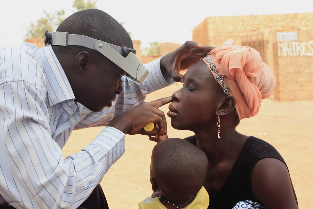
Photo: CDC / Unsplash
Ophthalmology documentation, from the exam lane to the OR
A comprehensive eye exam generates a lot of discrete data in a short visit. Corrected and uncorrected acuity for each eye, manifest refraction, pupils, motility, confrontation fields, tonometry, and then a slit lamp exam that walks the lids, conjunctiva, cornea, anterior chamber, iris, and lens before you even dilate. All of it has to land in the right field, per eye, and none of it is a place to guess.
Then the clinic mix shifts. A medical retina or glaucoma day is stacked with follow-ups where the story is in the trend: an IOP that crept from 18 to 24, a cup-to-disc ratio that looks a hair worse, a field defect deepening, or macular fluid on OCT that did or did not respond to the last anti-VEGF injection. Those notes lean on the numbers and your interpretation of the imaging, and they need to connect this visit to the last one.
Surgical work is a third shape. A cataract evaluation documents the lens grade, the visually significant complaint, biometry, and the discussion of lens options and risks. The one-day and one-week post-ops are quick but specific: pressure, the wound, the anterior chamber, the position of the implant. Transcribe Health listens to the visit or to your narration after a procedure and drafts the note in the shape that visit calls for. You read it and sign. Nothing is filed without you.
Documentation Challenges in Ophthalmology
- 1
Capturing dense, structured exam data (BCVA and refraction for each eye, IOP, pupils, slit lamp findings by segment, and dilated fundus) without turning the note into a data-entry chore
- 2
Switching between medical visits, surgical consults, and post-op checks in the same clinic session, where each note type has a different shape and a different level of detail
- 3
Documenting your read of OCT, visual fields, and fundus photos, plus in-office procedures like injections and lasers, in language that supports the code you bill
How Transcribe Health Helps Ophthalmology
Structured Exam Capture
Takes the visual acuities, IOP, slit lamp, and dilated fundus findings you call out and lays them into the note per eye, right and left, in the order you examine.
Imaging Interpretation Notes
Records the read you give on OCT, visual field, and fundus photos and drops it into the assessment, without touching the images themselves.
Procedure and Injection Documentation
Structures a note for an intravitreal injection, a laser, or a minor in-office procedure from your narration, including the eye, the agent, and consent.
An ophthalmology note, captured in real time
An abridged glaucoma follow-up, drafted the way you would see it before signing.
68-year-old woman returning for a 4-month glaucoma check. On latanoprost 0.005% one drop each eye at bedtime, reports good adherence and no ocular surface irritation. No new floaters, flashes, or vision changes. No headache or eye pain. Family history of glaucoma in her mother.
BCVA 20/25 right eye, 20/20 left eye. IOP by Goldmann applanation 17 mmHg right, 19 mmHg left, checked at 10:15 AM. Pupils equal and reactive, no APD. Slit lamp: lids and lashes normal, conjunctiva quiet, cornea clear, anterior chamber deep and quiet both eyes, mild nuclear sclerosis. Dilated fundus: C/D ratio 0.7 right eye, 0.75 left eye with mild inferior rim thinning on the left, maculae flat, vessels normal, periphery attached. OCT RNFL: borderline inferior thinning left eye, stable versus prior. Humphrey 24-2: superior nasal step left eye, unchanged from six months ago.
1) Primary open-angle glaucoma, both eyes, left worse than right, structurally and functionally stable on current therapy. 2) Left IOP 19 mmHg, slightly above target of high teens for this optic nerve. 3) Mild nuclear sclerotic cataract, both eyes, not visually significant.
Continue latanoprost 0.005% each eye at bedtime. Given the left pressure sitting at the upper edge of target and the inferior rim thinning, add timolol 0.5% one drop left eye twice daily and recheck IOP in 6 weeks. Repeat OCT RNFL and Humphrey 24-2 at the next visit to confirm stability. Reviewed drop technique and punctal occlusion. Return in 6 weeks for pressure check, sooner if any vision change.
Illustrative example. Not a real patient encounter.
Built for ophthalmology coding and your EMR
Notes capture the exam elements, the imaging you interpreted, and the medical decision-making that back up an eye visit or E/M level and a procedure's medical necessity. That detail is what payers look for, and the first thing to slip when you are running behind in clinic.
- 92004 / 92014Comprehensive eye exam, new or established patient
- 92134OCT of the retina, unilateral or bilateral
- 92083Visual field exam, extended (threshold, Humphrey 24-2 or 30-2)
- 67028Intravitreal injection of a pharmacologic agent
- 66984Cataract extraction with intraocular lens, one stage
- 92133OCT of the optic nerve, unilateral or bilateral
- H25.13Age-related nuclear cataract, bilateral
- H40.11X3Primary open-angle glaucoma, severe stage
- E11.359Type 2 diabetes with proliferative diabetic retinopathy without macular edema
- H35.31Nonexudative age-related macular degeneration
- H33.20Serous retinal detachment, unspecified eye
Fits the workflow you already have
Drafts drop into the EMRs ophthalmology practices actually run on, including Nextech, EyeMD EMR, and the ophthalmology-specific workflows in Epic. You get whatever note type you are working in, a comprehensive exam, a retina or glaucoma follow-up, or a pre-op consult. It writes up the OCT, visual field, and fundus reads you dictate. It does not open your imaging or replace your review of it; you interpret the scan and it records what you said.
Ophthalmology AI scribe questions
Does it handle per-eye exam data and ophthalmic terminology?
Can it document procedures, or only clinic exams?
Does it read my OCT or visual fields?
How is patient information protected?
Does it connect to our EMR?
Ready to Transform Your Documentation?
Join thousands of healthcare providers who save hours every day with AI-powered medical transcription.
Start Free Trial